










Understanding Progressive and Regressive Staining Methods
Entrance
Hematoxylin is the primary staining agent that forms the nuclear component of H&E staining. However, not all hematoxylins used in histopathology laboratories share the same formulation or staining approach.
Different hematoxylin formulations may vary in terms of oxidation system, mordant, excipients, and intended use. Additionally, there are two main approaches to controlling nuclear staining with hematoxylin: progressive and regressive.
Understanding these differences helps to understand why H&E staining results can vary between laboratories and protocols.
How does hematoxylin stain?
Hematoxylin is a naturally occurring compound that is converted into its active staining form during the staining process. Hematein, formed as a result of oxidation, forms complexes with suitable metal ions and binds to structures in the tissue, particularly those rich in nucleic acids.
Mordants and other components used in hematoxylin formulations can affect the resulting staining characteristics.
Therefore, “hematoxylin” should not be considered as a single standard product.
Types of hematoxylin
Different hematoxylin formulations are used in histopathology and cytology laboratories.
Commonly known formulations include:
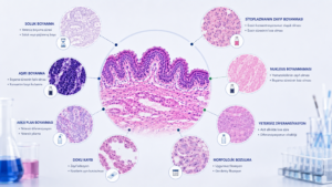
- Harris hematoxylin,
- Mayer hematoxylin,
- Gill hematoxylin,
- Ehrlich hematoxylin,
- Carazzi hematoxylin
Examples include these.
These formulations may have different intended uses and staining behaviors.
For example, some hematoxylins are more suitable for a progressive approach, while others can be used with a regressive staining approach.
Therefore, when evaluating a hematoxylin, one should look not only at its name but also at the product's formulation and recommended method of use.
What is progressive painting?
In progressive staining, tissue is stained with hematoxylin until the desired nuclear staining intensity is achieved.
A separate differentiation step may not be necessary for the controlled removal of excess paint later.
In this approach, staining time and hematoxylin properties are important. The aim is to ensure that the nuclei reach sufficient density while avoiding overstaining.
Some hematoxylins, such as Mayer and Gill, can be used with a progressive approach.
What is regressive painting?
In the regressive method, the tissue is initially stained at a level higher than the target intensity.
Excess hematoxylin is then removed through a controlled differentiation step.
The basic principle behind this method is:
Staining → Overstaining → Differentiation → Desired nuclear contrast
This can be summarized as follows.
In the regressive method, the duration and conditions of differentiation are very important. Insufficient differentiation can lead to excessively dark nuclei, while excessive differentiation can result in an undue reduction in nuclear staining.
Why is differentiation important?
Differentiation is one of the control points of the regressive hematoxylin method.
The goal is not to remove all of the hematoxylin, but to achieve adequate nuclear contrast by reducing the unwanted stain from the overstained tissue in a controlled manner.
Therefore, differentiation time does not have to be the same in every laboratory. Factors such as the hematoxylin used, tissue and section characteristics, and laboratory protocol should be taken into consideration.
What is bluing?
Nuclei stained with hematoxylin may appear reddish-purple during the staining process. Bluing ensures that the nuclear structures take on more characteristic blue-purple tones.
Bluing can be performed using water or suitable alkaline solutions.
This stage plays a crucial role in the final appearance of nuclear contrast.
The fundamental difference between progressive and regressive methods
| Feature | Progressive | Regressive |
| Painting approach | Painting to the desired intensity. | Over-staining followed by differentiation |
| Differentiation | A separate stage is usually not required. | It is one of the basic stages. |
| Checkpoint | Painting time and intensity | Painting + differentiation |
| Technical approach | More directly | It requires a more controlled intervention. |
| Protocol sensitivity | Depending on painting conditions. | Especially depending on the differentiation conditions. |
This table summarizes the key differences between the two methods. However, the specific hematoxylin formulation and manufacturer's protocol used in actual practice must be considered.
Which hematoxylin is better?
There is no single answer to this question.
Suitability of a hematoxylin:
- Depending on the type of tissue used,
- Regarding the painting method,
- Manual or automatic system,
- To achieve the desired nuclear contrast,
- According to the laboratory protocol,
- To the user's need for standardization,
- Compatibility with other painting stages
It may vary depending on the situation.
Therefore, just because a product dyes “darker” or “faster” doesn’t automatically mean it’s better.
In routine histopathology, it is important to obtain reliable and reproducible nuclear staining.
Standardization in hematoxylin selection.
One of the key considerations in hematoxylin selection is standardization.
The nuclear staining characteristics may change when the formulation of hematoxylin or the working conditions used in the same laboratory change.
In laboratories where a large number of preparations are evaluated, it is important to standardize the staining protocol within certain limits.
Why is this important for H&E?
Hematoxylin is only one component of H&E staining. However, it plays a critical role in the contrast of nuclei and the evaluation of morphological details.
Therefore, the choice of hematoxylin is as important as:
- Painting time,
- Differentiation,
- Bluing,
- The contrast created with eosin,
- Section quality,
- Tissue processing conditions
They should be evaluated together.
Conclusion
Hematoxylins can have different formulations and different staining behaviors. Progressive and regressive staining approaches are two fundamental methods in controlling nuclear staining.
In the progressive method, the tissue is stained until the desired density is reached, while in the regressive method, excessive staining is performed, and then some of the unwanted stain is removed by differentiation.
The goal of both methods is the same: to obtain sufficient, balanced, and reproducible nuclear contrast for microscopic evaluation.
Therefore, hematoxylin selection should not be based on "the product that produces the darkest stain,", laboratory needs, protocol and reproducibility objectives It should be evaluated based on this.
To inform
This content has been prepared for general informational purposes, drawing on scientific literature and reliable technical sources regarding hematoxylin formulations and staining approaches. For specific applications, the technical documentation of the product used and the validated procedures of the laboratory should be considered.